

|
Product Details:
|
|
| Brand Name: | ALOKA |
|---|---|
| Model Number: | UST-9120 |
|
Payment & Shipping Terms:
|
|
| Minimum Order Quantity: | 1 |
| Price: | Negotiable |
| Packaging Details: | Neutral Package |
| Delivery Time: | 1-3 work days |
| Payment Terms: | Western Union, T/T |
|
Detail Information |
|||
| Category: | Supersonic Sensor | Description: | Radiography Scanner |
|---|---|---|---|
| Conditon: | Good | Purpose: | Equipment Used In Hospitals |
| Warranty: | 60 Days/ 90 Days | Service: | Outright Sale |
| High Light: | Hospital Ultrasound Scanner Probe,Aloka UST 9120 Ultrasound Scanner Probe |
||
Product Description
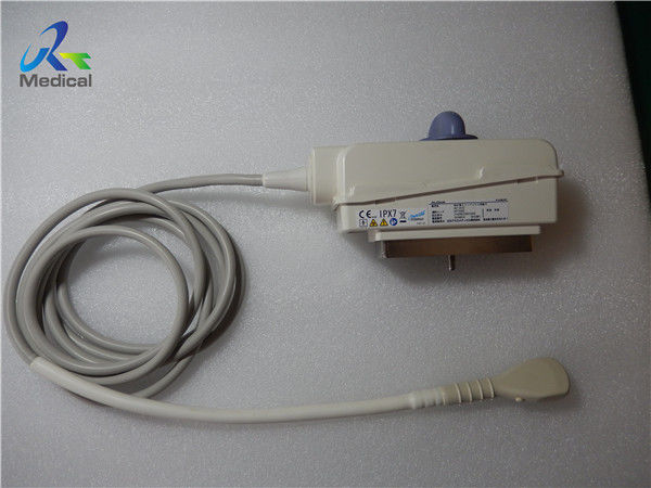
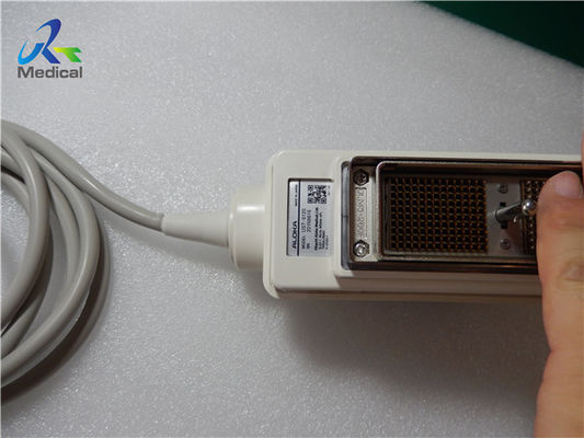
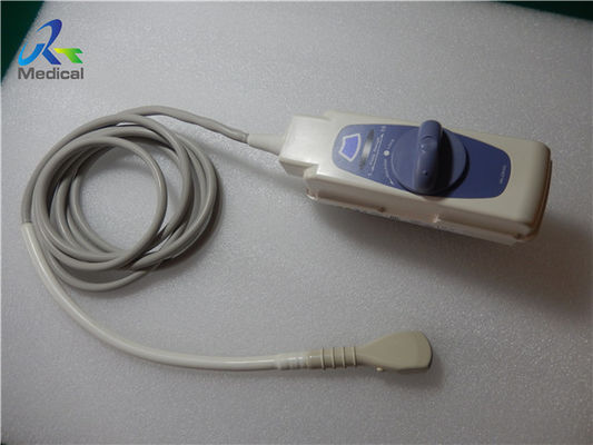
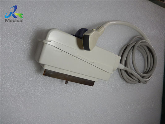
Medical Ultrasound Transducer Probe Aloka UST-9120/Convex Array/Used In Hospital
1. Type: Convex Array
2. Compatible system: Aloka Alpha 5, Alpha 7, Alpha 10
3. Application:Small Parts, Neonatal
4. Condition: original, in good working condition
6. With 60 days warranty
![]()
![]()
Parts of Aloka probes we can offer:
| Brand | Model | Compatible System |
| Aloka | UST-979-3.5 | SSD-900/1000/1100/1400/1700/3500/4000 |
| Aloka | UST-944B-3.5 | SSD-500/SSD- 620/SSD- 625/SSD-650/SSD-1100 |
| Aloka | UST-669-5-7.5 | SSD-1000/ SSD -1700/ SSD -2200 |
| Aloka | UST-984-5 | SSD-1000/SSD-1400/SSD-1700/SSD-5000/SSD-5500 |
| Aloka | UST-9115-5 | SSD-5000/ Alpha 5/ Alpha 10 |
| Aloka | UST-9116P-5 | SSD-1000 |
| Aloka | UST-9118 | Alpha 5/Alpha 10/SSD-3500/SSD-4000/SSD-5500 |
| Aloka | UST-9120 | Alpha 5/Alpha 7/Alpha 10 |
| Aloka | UST-9121 | SSD-3500/SSD-4000 |
| Aloka | UST-9123 | SSD-4000/ SSD-3500 |
| Aloka | UST-9124 | SSD-3500/SSD-4000 |
| Hitachi-Aloka Ultrasound System |
| F31, F37, SSD-3500, SSD-3500 SV, SSD-4000, SSD-5000, ALPHA 5, ALPHA 6, ALPHA 7, ALPHA 10, PROSOUND F75, HI VISION AVIUS, HI VISION PREIRUS, EUB-5500, EUB-6500, EUB-7500, EUB-8500, Arietta 60, Arietta 70, Ascendus... |
Knowledge point
Color-flow imaging
Color-flow imaging is an enhanced form of Doppler ultrasound technology. In a procedure similar to duplex ultrasound, it uses color to highlight the direction of blood flow. Vessels in which blood is flowing are colored red for flow in one direction and blue for flow in the other, with a color scale that reflects the speed of the flow.It can clearly understand the anatomical shape and activity of large blood vessels, and can visually display the direction, speed, range of blood flow, and whether there are blood flow disorders and abnormal pathways. The speed of blood flow determines the reflection frequency, which is represented by amplitude beam in spectral Doppler. The velocity of blood flow is fast, and the amplitude on the spectrum curve is high; the velocity of blood flow is slow, and the amplitude on the spectrum curve is low, so amplitude can accurately calculate the velocity of blood flow. Color Doppler images are displayed with different brilliance.
3D
In the ultrasonic detector, the detected 3Dobject image is displayed as a 3Ddisplay method by plane display. Three dimensional reconstruction refers to the use of ultra wide-band technology, on the basis of a large number of high-definition two-dimensional image accurate data provided, to make the collected image signal data characteristic and systematic, so as to form a 3Ddisplay. Its unique control signal function will make a series of 3Dimages fully displayed on the screen.
3D image reconstruction
3D image reconstruction is a hot spot in ultrasound image processing and has become a development trend of ultrasound imaging. The first 3Dultrasound imaging commercial device uses mechanical scanning probes that oscillate in perpendicular directions to collect data of interest within 3 seconds, and perform image reconstruction to generate sagittal, coronal, and cross-sectional images. These planes can be adjusted within the range of ultrasound information capacity, and multiple continuous images can be seen.
There are many problems to be solved in 3Dultrasound imaging, including data acquisition methods, real-time image reconstruction, and clinical reference value. At present, four data collection methods have emerged: parallel scanning, rotating scanning, sector scanning, and free-hand Scanning. The most eye-catching 3Dultrasound imaging is real-time 3Dimaging. The key to real-time 3Dimaging is to use parallel data processing and shorten data acquisition time. Acoustic pulses are emited in several directions at the same time, and simultaneously acquires and processes multiple sound beam information of the scan line, which obviously increases the complexity of the ultrasound imaging system.
Enter Your Message
| Guangzhou Rongtao Medical Tech LTD. |
| Room 1705,Office Building B1,Wanda Plaza,No.2707,Kaichuang Road,Guangzhou,Guangdong,China |
| miley@ultrasound-service.com |