

|
Product Details:
|
|
| Brand Name: | ALOKA |
|---|---|
| Model Number: | UST-9121 |
|
Payment & Shipping Terms:
|
|
| Minimum Order Quantity: | 1 |
| Price: | Negotiable |
| Packaging Details: | Neutral Package |
| Delivery Time: | 1-3 work days |
| Payment Terms: | Western Union, T/T |
|
Detail Information |
|||
| Category: | Supersonic Sensor | Description: | Medical And Hospital Supplies |
|---|---|---|---|
| Conditon: | Original Probe | Purpose: | Equipment Used In Hospitals |
| Service: | Outright Sale | Type: | Medical Doppler |
| Packing: | 1pcs.box | ||
| High Light: | Tight Convex Ultrasound Scanner Probe,14mm Ultrasound Scanner Pro |
||
Product Description
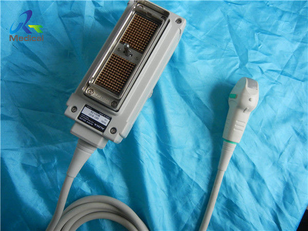
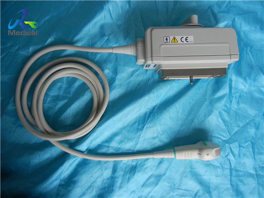
Aloka UST-9121 14mm Multi Frequency Tight Convex Transducer
![]()
![]()
Parts of Aloka probes we can offer:
| Brand | Model | Compatible System |
| Aloka | UST-979-3.5 | SSD-900/1000/1100/1400/1700/3500/4000 |
| Aloka | UST-944B-3.5 | SSD-500/SSD- 620/SSD- 625/SSD-650/SSD-1100 |
| Aloka | UST-669-5-7.5 | SSD-1000/ SSD -1700/ SSD -2200 |
| Aloka | UST-984-5 | SSD-1000/SSD-1400/SSD-1700/SSD-5000/SSD-5500 |
| Aloka | UST-9115-5 | SSD-5000/ Alpha 5/ Alpha 10 |
| Aloka | UST-9116P-5 | SSD-1000 |
| Aloka | UST-9118 | Alpha 5/Alpha 10/SSD-3500/SSD-4000/SSD-5500 |
| Aloka | UST-9120 | Alpha 5/Alpha 7/Alpha 10 |
| Aloka | UST-9121 | SSD-3500/SSD-4000 |
| Aloka | UST-9123 | SSD-4000/ SSD-3500 |
| Aloka | UST-9124 | SSD-3500/SSD-4000 |
Knowledge point
Color Doppler flow imaging
Color Doppler flow imaging system can simultaneously display B-type image and Doppler blood flow data (blood flow direction, flow velocity, flow dispersion) of dual ultrasound scanning system. Color power angio(CPA) detected the backscattered energy of blood cells in the blood flow, which did not distinguish the flow direction, and had nothing to do with the angle θ (the angle between the direction of sound wave and the direction of blood flow). CPA can improve the sensitivity of blood flow detection, especially suitable for displaying low-speed blood flow of small vessels, but can not show the direction of blood flow.
Harmonic imaging
Harmonic imaging exploits non-linear propagation of ultrasound through the body tissues. When the frequency of emitted sound wave is f 0, the frequency of echo (due to reflection or scattering) includes f 0 (called fundamental wave), 2f 0, 3F 0 and so on The second harmonic (2f 0) has the largest energy.
Ultrasonic harmonic imaging (UHI) is based on the information of human body carried by the second harmonic in echo (reflection or scattering). Harmonic imaging without UCA is called native harmonic imaging or tissue harmonic imaging. Harmonic imaging using UCA (ultrasound contrast agent) is called contrast harmonic imaging.
Multi-frequency probe
Multi-frequency probe is a new development of pulse-echo transducer. The probe can send out several different ultrasonic pulses, so as to cover the near field with high-frequency ultrasound, the intermediate-frequency ultrasound to cover the transition zone between the far and near fields, and the low-frequency ultrasound to cover the far field. The unit multi-frequency probe is to bond multiple layers of piezoelectric ceramics (or polymer piezoelectric materials) to each other, and lead wires from the electrodes between each layer to obtain multiple frequency ultrasonic pulse emission through exciting different layers . The digital coding of the multi-frequency probe is simple but easy to lose the signal.
Enter Your Message
| Guangzhou Rongtao Medical Tech LTD. |
| Room 1705,Office Building B1,Wanda Plaza,No.2707,Kaichuang Road,Guangzhou,Guangdong,China |
| miley@ultrasound-service.com |