

|
Product Details:
|
|
| Brand Name: | ALOKA |
|---|---|
| Model Number: | UST-5543 |
|
Payment & Shipping Terms:
|
|
| Minimum Order Quantity: | 1 |
| Price: | Negotiable |
| Packaging Details: | Neutral Package |
| Delivery Time: | 1-3 work days |
| Payment Terms: | Western Union, T/T |
|
Detail Information |
|||
| Category: | Medical Facility | Description: | Medical Instruments Suppliers |
|---|---|---|---|
| Conditon: | Original Probe | Purpose: | Medical Apparatus |
| Application: | Small Parts | Frequency: | 5-10 MHz |
| High Light: | UST 5543 Ultrasound Scanner Probe,Alpha 10 system Ultrasound Scanner Probe |
||
Product Description
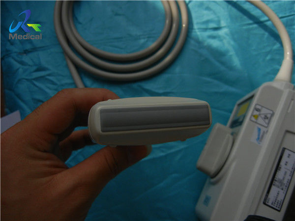
Aloka UST-5543 Linear Small Parts Transducer For Aloka Alpha 5/Alpha 10
1. Type:linear array
2. Frequency: 5-10 MHz
3. Compatible system: Aloka Alpha 5/Alpha 10
4. Application: small parts
5. Condition: new, original, in excellent working condition
![]()
Other Aloka Probes we can offer:
| Brand | Model | Compatible System |
| Aloka | UST-979-3.5 | SSD-900/1000/1100/1400/1700/3500/4000 |
| Aloka | UST-944B-3.5 | SSD-500/SSD- 620/SSD- 625/SSD-650/SSD-1100 |
| Aloka | UST-669-5-7.5 | SSD-1000/ SSD -1700/ SSD -2200 |
| Aloka | UST-984-5 | SSD-1000/SSD-1400/SSD-1700/SSD-5000/SSD-5500 |
| Aloka | UST-9115-5 | SSD-5000/ Alpha 5/ Alpha 10 |
| Aloka | UST-9116P-5 | SSD-1000 |
| Aloka | UST-9118 | Alpha 5/Alpha 10/SSD-3500/SSD-4000/SSD-5500 |
| Aloka | UST-9120 | Alpha 5/Alpha 7/Alpha 10 |
| Aloka | UST-9121 | SSD-3500/SSD-4000 |
| Aloka | UST-9123 | SSD-4000/ SSD-3500 |
| Aloka | UST-9124 | SSD-3500/SSD-4000 |
Knowledge point
Multi-frequency probe
Multi-frequency probe is a new development of pulse-echo transducer. The probe can send out several different ultrasonic pulses, so as to cover the near field with high-frequency ultrasound, the intermediate-frequency ultrasound to cover the transition zone between the far and near fields, and the low-frequency ultrasound to cover the far field. The unit multi-frequency probe is to bond multiple layers of piezoelectric ceramics (or polymer piezoelectric materials) to each other, and lead wires from the electrodes between each layer to obtain multiple frequency ultrasonic pulse emission through exciting different layers . The digital coding of the multi-frequency probe is simple but easy to lose the signal.
![]()
Wide-band probe
The wide-band probe send out continuous ultrasonic pulse signals, so that the ultrasonic signals in a certain frequency range can be transmitted and received without gaps.
Ultra-wideband probe
On the basis of the wide-band probe, the ultrasonic signal range that the probe receives and transmits is further expanded. The signal of the ultra-wideband probe is completely carried out at the moment of reception, and is fully digitally coded and amplified to ensure that the signal is not distorted and expand the dynamic range of the signal.
Annular probe
The principle of using the circular ring array dynamic segmented focusing method in mechanical sector-scan ultrasonic diagnostic equipment is the same as that of linear array dynamic focusing. Applying a proper delay to the electrical signal of each element can achieve focusing at any distance along the central axis, which is similar to the function of an acoustic lens, so it functions as an "electronic focusing lens".
Color-flow imaging
Color-flow imaging is an enhanced form of Doppler ultrasound technology. In a procedure similar to duplex ultrasound, it uses color to highlight the direction of blood flow. Vessels in which blood is flowing are colored red for flow in one direction and blue for flow in the other, with a color scale that reflects the speed of the flow.It can clearly understand the anatomical shape and activity of large blood vessels, and can visually display the direction, speed, range of blood flow, and whether there are blood flow disorders and abnormal pathways. The speed of blood flow determines the reflection frequency, which is represented by amplitude beam in spectral Doppler. The velocity of blood flow is fast, and the amplitude on the spectrum curve is high; the velocity of blood flow is slow, and the amplitude on the spectrum curve is low, so amplitude can accurately calculate the velocity of blood flow. Color Doppler images are displayed with different brilliance.
Enter Your Message
| Guangzhou Rongtao Medical Tech LTD. |
| Room 1705,Office Building B1,Wanda Plaza,No.2707,Kaichuang Road,Guangzhou,Guangdong,China |
| miley@ultrasound-service.com |