

|
Product Details:
|
|
| Brand Name: | ALOKA |
|---|---|
| Model Number: | UST-5548 |
|
Payment & Shipping Terms:
|
|
| Minimum Order Quantity: | 1 |
| Price: | Negotiable |
| Packaging Details: | Neutral Package |
| Delivery Time: | 1-3 work days |
| Payment Terms: | Western Union, T/T |
|
Detail Information |
|||
| Category: | Imaging Center Facility | Description: | Imaging Center Device |
|---|---|---|---|
| Conditon: | Original Hitachi Aloka | Purpose: | Medical Apparatus |
| Application: | Vascular Probe | Frequency: | 3.0-7.5 Mhz |
| High Light: | Linear Peripheral Ultrasound Scanner Probe,42mm Ultrasound Scanner Probe,42mm vascular probe ultrasound |
||
Product Description
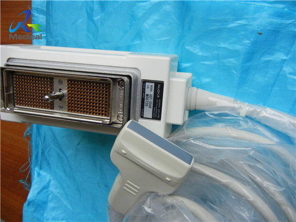
Aloka UST-5548 42mm Multi Frequency Linear Peripheral Vascular Transducer
1. Type:linear
2. Frequency: 3.0-7.5 Mhz
3. Compatible system: Alpha 5SX/Alpha 7/Alpha 10/SSD-3500SX
4. Application: vascular
5. Condition: original, in good working condition
6. With 60 days warranty
![]()
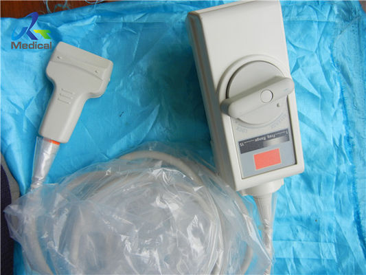
Other Aloka Probes we can offer:
| Brand | Model | Compatible System |
| Aloka | UST-979-3.5 | SSD-900/1000/1100/1400/1700/3500/4000 |
| Aloka | UST-944B-3.5 | SSD-500/SSD- 620/SSD- 625/SSD-650/SSD-1100 |
| Aloka | UST-669-5-7.5 | SSD-1000/ SSD -1700/ SSD -2200 |
| Aloka | UST-984-5 | SSD-1000/SSD-1400/SSD-1700/SSD-5000/SSD-5500 |
| Aloka | UST-9115-5 | SSD-5000/ Alpha 5/ Alpha 10 |
| Aloka | UST-9116P-5 | SSD-1000 |
| Aloka | UST-9118 | Alpha 5/Alpha 10/SSD-3500/SSD-4000/SSD-5500 |
| Aloka | UST-9120 | Alpha 5/Alpha 7/Alpha 10 |
| Aloka | UST-9121 | SSD-3500/SSD-4000 |
| Aloka | UST-9123 | SSD-4000/ SSD-3500 |
| Aloka | UST-9124 | SSD-3500/SSD-4000 |
Knowledge point
3D
In the ultrasonic detector, the detected 3Dobject image is displayed as a 3Ddisplay method by plane display. Three dimensional reconstruction refers to the use of ultra wide-band technology, on the basis of a large number of high-definition two-dimensional image accurate data provided, to make the collected image signal data characteristic and systematic, so as to form a 3Ddisplay. Its unique control signal function will make a series of 3Dimages fully displayed on the screen.
3D image reconstruction
3D image reconstruction is a hot spot in ultrasound image processing and has become a development trend of ultrasound imaging. The first 3Dultrasound imaging commercial device uses mechanical scanning probes that oscillate in perpendicular directions to collect data of interest within 3 seconds, and perform image reconstruction to generate sagittal, coronal, and cross-sectional images. These planes can be adjusted within the range of ultrasound information capacity, and multiple continuous images can be seen.
There are many problems to be solved in 3Dultrasound imaging, including data acquisition methods, real-time image reconstruction, and clinical reference value. At present, four data collection methods have emerged: parallel scanning, rotating scanning, sector scanning, and free-hand Scanning. The most eye-catching 3Dultrasound imaging is real-time 3Dimaging. The key to real-time 3Dimaging is to use parallel data processing and shorten data acquisition time. Acoustic pulses are emited in several directions at the same time, and simultaneously acquires and processes multiple sound beam information of the scan line, which obviously increases the complexity of the ultrasound imaging system.
Color Doppler flow imaging
Color Doppler flow imaging system can simultaneously display B-type image and Doppler blood flow data (blood flow direction, flow velocity, flow dispersion) of dual ultrasound scanning system. Color power angio(CPA) detected the backscattered energy of blood cells in the blood flow, which did not distinguish the flow direction, and had nothing to do with the angle θ (the angle between the direction of sound wave and the direction of blood flow). CPA can improve the sensitivity of blood flow detection, especially suitable for displaying low-speed blood flow of small vessels, but can not show the direction of blood flow.
Harmonic imaging
Harmonic imaging exploits non-linear propagation of ultrasound through the body tissues. When the frequency of emitted sound wave is f 0, the frequency of echo (due to reflection or scattering) includes f 0 (called fundamental wave), 2f 0, 3F 0 and so on The second harmonic (2f 0) has the largest energy.
Ultrasonic harmonic imaging (UHI) is based on the information of human body carried by the second harmonic in echo (reflection or scattering). Harmonic imaging without UCA is called native harmonic imaging or tissue harmonic imaging. Harmonic imaging using UCA (ultrasound contrast agent) is called contrast harmonic imaging.
Enter Your Message
| Guangzhou Rongtao Medical Tech LTD. |
| Room 1705,Office Building B1,Wanda Plaza,No.2707,Kaichuang Road,Guangzhou,Guangdong,China |
| miley@ultrasound-service.com |