

|
Product Details:
|
|
| Brand Name: | GE |
|---|---|
| Model Number: | 4C |
|
Payment & Shipping Terms:
|
|
| Minimum Order Quantity: | 1 |
| Price: | Negotiable |
| Packaging Details: | Neutral Package |
| Delivery Time: | 1-3 work days |
| Payment Terms: | T/T, Western Union |
|
Detail Information |
|||
| Description: | Diagnosis Machine | Category: | Scan Image |
|---|---|---|---|
| Warranty: | 60 Days | Service: | Outright Sale |
| Item: | Diagnostic Tools | Condition: | Original GE |
| High Light: | Convex Array Ultrasound Transducer Probe,GE 4C Ultrasound Transducer Probe |
||
Product Description
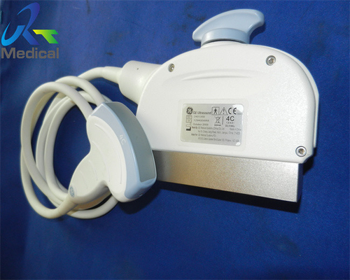
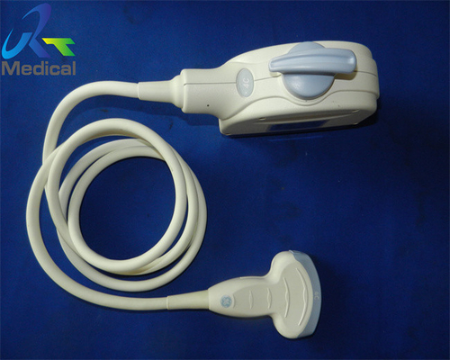
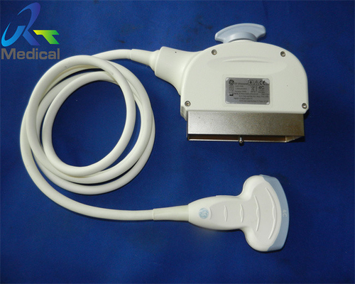
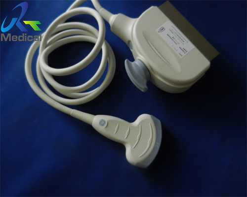
GE 4C Convex Array Ultrasound Transducer Used Probe Use in Hospital Diagnosis Machine
1.Type: convex array
2.Frequency:1.5-4.5 MHz
3.Compatible system: Logiq & Vivid Series
4.Application: OB/GYN, abdominal, vascular and urology
5.Condition: pre-owned one, original, in good working condition
6.With 60 days warranty
![]()
![]()
![]()
Parts of GE probes we can offer:
| Brand | Model | Compatible System |
| GE | 3.5C | Logiq 3/ Logiq 5/ Logiq 7/ Logiq 9/ Logiq A5/ Logiq P5/ Logiq S6/Vivid7 |
| GE | 3C | Logiq 3/ Logiq 5 |
| GE | 3Cb | Logic 200 Pro |
| GE | 3CRF | Logiq S6 |
| GE | 3CRF-D | Logiq S8/ Logiq E9 |
| GE | 3SP | LOGIQ P |
| GE | 3Sp-D | Voluson E6/Voluson E8 / Bt08 & Higher version |
| GE | 3S | vivid & Logiq series |
| GE | 3S-RS | Loqigbook/XP/ Vivid-I |
| GE | 3S-SC | venue 40 |
| GE | 3S-RC | Logiq C5/ Logiq C2/ Logiq C3 |
| GE | 3SC-RS | Vivid S5/ Vivid iq/ Vivid i |
| GE | 3.8CA | Logiq P3 |
| GE | 3V-D | Vivid E9 |
| GE | 3.8C-RC | Loqig C2/Logiq C3/ Logiq C5/ Logiq premiums |
| GE | 4C | Logiq & Vivid Series |
| GE | 4C-RC | Logiq C2/Logiq C3/Logiq C5/Logiq Premium |
| GE | 4C-RS | Vivid i Square/ Logiq e/ Vivid e |
| GE | 4C-SC | Venue 40 |
| GE | 4C-A | Voluson 730 |
| GE | 4C-D | Voluson E6/ E8 and Vivid E9 |
| GE | 4D3C-L | Logiq 9/ Logiq A5/ Logiq P5/ Logiq S6 |
| GE | 4V-D | Vivid E9 |
| GE | 4S | Logiq and Vivid series |
| GE | 5S | pediatric cardiac |
| GE | 5S-RS | Vivid q/ Vivid i/Vivid S5/Vivid S6 |
| GE | 6.5C-RC | Vivid q/ Vivid i |
| GE | 7L | Logiq and Vivid series |
| GE | 7L-RC | Logiq C2/ C5/ Logiq Premium |
| GE | 739L | Logiq 500 Pro/Vivid 3/Logiq 400 MD/Logiq 500 MD |
| GE | 7S | Logiq9/ Logiq 7/ Logiq S6/ Logiq 5/ Logiq 3 /Vivid 7/Vivid 3 |
| GE | 7.5L-RC | Logiq C2/Logiq C3/Logiq C5/Logiq Premium |
| GE | 8C | Logiq & Vivid series |
| GE | 8C-RS | Voluson i/ Voluson e/ Logiq i/ Logiqbook XP/ Logiqbook/ Vivid i (from rev. 6.1.0)/ Vivid S5 |
| GE | 8L-RS | Logiq e/ Vivid e/ Vivid i and Logiq Book XP |
| GE | 9L | Vivid 7 Pro/Vivid 3/Logiq 9/ Logiq 7/Logiq 5/Logiq 3/ Logiq 400 |
| GE | 9L-D | Voluson E6/Voluson E8/Vivid E9 |
| GE | 9L-RS | logiq e/ vivid e/ vivid I/ vivid q/ vivid s5/ vivid s6 |
| GE | 10Lb-RS | Logiq book |
| GE | 10S-RS | Logiq Book/ Logiq e/ Vivid e/ Vivid i/ Logiq i/ Vivid i Square |
| GE | 10L | Logiq 3/5/7/9/ Vivid 3/4/A5/P5/7 Expert/7 Pro/ Logiq S6 |
| GE | 11L | Logiq P5/A5/A5Pro/ Logiq P6/P6 Pro |
| GE | 11L-D | premium GE ultrasound machines |
| GE | 12L | Logiq & Vivid series |
| GE | 12L-RS | Logiq e/ Logiq l/ Vivid e/Vivid I Voluson e/Voluson I/Vivid Q/Vivid S5/ Vivid S6 |
| GE | 12L-SC | Venue 40 |
| GE | 12S-D | Vivid E9 |
| GE | AB2-7 | Voluson S6/ Voluson S8/Voluson 730 |
| GE | AC2-5 | Voluson 730 |
| GE | BE9C | Logiq series |
Knowledge Point
Color Doppler flow imaging
Color Doppler flow imaging system can simultaneously display B-type image and Doppler blood flow data (blood flow direction, flow velocity, flow dispersion) of dual ultrasound scanning system. Color power angio(CPA) detected the backscattered energy of blood cells in the blood flow, which did not distinguish the flow direction, and had nothing to do with the angle θ (the angle between the direction of sound wave and the direction of blood flow). CPA can improve the sensitivity of blood flow detection, especially suitable for displaying low-speed blood flow of small vessels, but can not show the direction of blood flow.
Harmonic imaging
Harmonic imaging exploits non-linear propagation of ultrasound through the body tissues. When the frequency of emitted sound wave is f 0, the frequency of echo (due to reflection or scattering) includes f 0 (called fundamental wave), 2f 0, 3F 0 and so on The second harmonic (2f 0) has the largest energy.
Ultrasonic harmonic imaging (UHI) is based on the information of human body carried by the second harmonic in echo (reflection or scattering). Harmonic imaging without UCA is called native harmonic imaging or tissue harmonic imaging. Harmonic imaging using UCA (ultrasound contrast agent) is called contrast harmonic imaging.
Enter Your Message
| Guangzhou Rongtao Medical Tech LTD. |
| Room 1705,Office Building B1,Wanda Plaza,No.2707,Kaichuang Road,Guangzhou,Guangdong,China |
| miley@ultrasound-service.com |

